Imaging
Digital Radiographs
Willow Pet Hospital has the latest modern digital radiograph (x-ray) equipment.
Fast
This modern technology provides the best digital images available in just seconds. This facilitates less time that the patient must be on the table while taking images and therefore creates less stress and anxiety for the pet.
If image quality is affected by incorrect positioning, technique or patient movement, images can be retaken in a matter of seconds.
Highest Quality
The image quality compared to old film radiographs is truly unbelievable. Detail is what matters when talking about radiographs, this allows us to obtain a correct diagnosis and appropriate treatment.
Value
Some common uses for radiographs that help us define the best treatment and care for your pet:
• Coughing – Congestive Heart Failure, Pneumonia, Asthma, Tracheal Collapse, Megaesophagus
• Vomiting – ingestion of foreign
material, tumors/cancer
• Inappropriate urination – bladder/kidney stones, enlarged kidneys
• Lameness – fractures, ACL injuries, hip dysplasia, shoulder and elbow injuries, cancer, fungal infections, and tendonitis
• Paresis (weakness)/Paralysis
– intervertebral disc disease “slipped disc”, arthritis
Ultrasound
Willow Pet Hospital has the latest modern Esaote ultrasound equipment.
Value
Some common uses for ultrasound that help us define the best treatment and care for your pet:
• FAST Scans – brief, four quadrant scans of the abdomen for patients with suspected abdominal fluid from such
disease like a bleeding spleen, ruptured bladder or after a severe injury like being hit by a car
• Echocardiogram – an ultrasound of the heart essential for diagnosis of congestive heart failure (CHF) and the source of murmurs
(additional sounds from the heart)
• Ultrasound Guided Cystocentesis – guided sampling of urine directly from the bladder with the added benefit of evaluating the bladder for abnormalities including stones or cancer
•
Abdominal Ultrasound – evaluation of the liver, gall bladder, spleen, kidneys, bladder and intestines for abnormalities. Guided biopsies can also be taken during this procedure.
Expert Opinions
With a click of the button we can send these digital radiographs and ultrasound images to a board-certified radiologist for a second opinion. Radiologist have four additional years of specialized training, take an intensive
exam in their specialty and are required to maintain an advance knowledge in their specialty. Beyond the training they are focused on one thing all day long – radiology and ultrasound. That added intense experience really pays off. Additionally,
with an echocardiogram we can send these ultrasound images to the University of Minnesota and have a consultation with a cardiologist and get an expert opinion from a veterinary heart specialist.
Fleas & Ticks
Fleas
Fleas are the most common external parasite of companion animals. They live by consuming the blood of their hosts. Adults fleas are about 3 mm long and brown in color. Their bodies are flattened sideways which enables them to easily move through their host’s fur and they have strong claws that prevent them from being dislodged. They also have hind legs that are adapted for jumping which allows them to leap up to 50 times their body length. Stray cats and dogs, raccoons and rabbits can carry flea eggs into your yard. Fleas can survive in lows as cold as 28F and highs up to 95F.
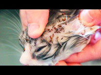
Many pet owners are surprised to learn their pet has fleas because they have not noticed them. In many cases we do not see the fleas until a thorough exam is performed. This is because many animals lick, groom and chew after being bitten by a flea, causing the flea to either jump off, or get swallowed by the pet.
Fleas are dangerous because they can cause a number of health problems with our pets including:
• Anemia (low red blood cell count in the body) due to blood loss. This anemia can be life threatening in smaller or very young animals and even
in larger animals, if there is a heavy flea infestation present.
• Severe itching which leads to hair loss and secondary skin infections. In fact, some animals are allergic to flea bites and they will develop a condition called flea allergy
dermatitis. This is commonly found in dogs and cats and can be very uncomfortable and requires medical treatment to resolve.
• A bacterial infection called Bartonella that is transmitted when they bite. This not only causes health issues in
dogs and cats but can affect people as well.
• A type of intestinal tapeworm called Dipylidium caninum can be transmitted if a dog or cat ingests the flea. The flea body contains an immature form of the parasite and if ingested will mature
in the intestine of the dog or cat and compete for the nutrients of the pet.
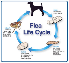
The flea life cycle consists of egg, larval, pupal, and adult stages.
1) Eggs are laid in the hair coat and fall off your pet into your home.
2) Larvae hatch from the eggs and develop in a pet’s environment by feeding on adult flea feces
(i.e. digested blood) that falls out of the hair coat of the pet.
3) Larvae eventually spin cocoons, often within carpet fibers, for pupation. The pupae in the cocoons are resistant to freezing, drying, and insecticides, and can lie dormant
for many months!
4) New fleas develop from pupae and can begin feeding within hours of finding a dog or cat. The entire flea life cycle can be completed in as little as three weeks.
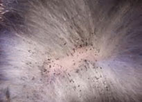
We diagnose a flea infestation by either seeing adult fleas or by using a flea comb and pulling flea “dirt”. Flea dirt is actually flea fecal material. Fleas take blood meals of your pet and their fecal material contains blood. If we rinse flea dirt with a small amount of water on a white paper towel, the spots will turn a rusty brown.
Fleas are very difficult problem to solve and certainly prevention is much less expensive and stressful than treatment. One female adult flea produces 40 to 50 eggs per day. You can see how quickly this can get out of control. The other issue is that only about 1% of the population of fleas, eggs and larvae live on your pet and the other 99% live in the environment. That is to say, they exist in your homes carpet, bedding, couches, clothing, etc. This makes treatment very frustrating.
Treatment includes treating EVERY pet in the house (dogs and cats) for ideally 6 months, at a minimum of 3 months, with an oral or topical flea and tick medication. Products we carry for dogs include: Frontline Gold topical, Nexgard oral and Bravecto oral medications. Products we carry for cats include: Frontline Gold topical, Revolution topical and Bravecto topical.
Treatment also includes aggressively treating the environment. This may take multiple treatments and includes: spot sprays (i.e. Knockout) and room foggers (i.e. Adams or Raid) and washing all bedding in areas where the pet sleeps. We have Knockout
spray and Adams room fogger products available.
It is also recommended to vacuum all floors multiple times and then replace the bag or wash out the vacuum canister thoroughly. Remember, fleas thrive particularly well in the well-regulated
temperatures in the home and love to develop in carpeting and the cracks between the boards of hard wood floors.
It may take multiple repeated treatments to eliminate the fleas and their entire life cycle of larvae and eggs. By the time you
see active fleas on your pet they have been living in your home for at least 2 months.
Of course, after this infestation has been resolved, we recommend monthly flea/tick preventative for all your pets.
Ticks
Ticks are external parasites that feed on the blood of their hosts. They are attracted to motion, body heat, and carbon dioxide exhaled by mammals making people, dogs, cats and other mammals their ideal hosts.
The two most common ticks we have in this area are the American Dog tick and the Deer tick. They are slightly different in appearance and the Deer ticks are smaller than the Dog ticks.
Deer ticks are the ticks responsible for transmitting diseases in our area.
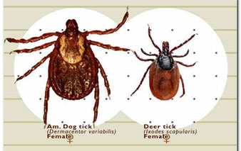
Deer ticks attach to the host to take a blood meal and in the process of taking this meal they can transmit bacterial diseases to the host. The nymph Deer ticks are almost microscopic in size so you may never know your dog was ever bitten by one.
Anaplasmosis and Lyme disease are the two most common tick diseases that we encounter in our area. These tick diseases cause potentially life-threatening symptoms and cause your pet pain and suffering.
Some symptoms of Lyme disease include:
• Fever
• Lethargy
• Stiffness and reluctance to move
• Swollen joints
• Shifting leg lameness
• Enlarged lymph nodes
• Rarely neurologic signs are noted
• The most serious
potential consequence of Lyme disease is a type of kidney damage that leads to acute and progressive kidney failure.
Some symptoms of Anaplasmosis include:
• Fever
• Lethargy
• Stiffness and reluctance to move
• Swollen joints
• Shifting leg lameness
• Enlarged lymph nodes
• Signs of uncontrolled internal bleeding, such as bloody nose,
bruising, and dark blood in the stool.
We recommend protecting your pet in multiple ways:
• TESTING FOR EXPOSURE: Screening dogs yearly for exposure to Lyme and Anaplasmosis organisms is important. We do this by using a simple in-house combination heartworm/tick disease screening
blood test.
• VACCINATION: Vaccination against Lyme disease to allow your pet to develop immunity against the bacteria responsible for causing this disease. This vaccine requires 2 initial boosters 2 to 3 weeks apart and is then repeated annually.
• FLEA/TICK PREVENTATIVES: To reduce the risk of transmission of other tick diseases to your pet. We have both topical and oral flea and tick preventative products available.
Dentisty
Dental Care
Our Values – Dental Care
The mouth and teeth are critical components to a dog and cat’s quality of life. Unfortunately, it is also one of the most overlooked. It is our view that dogs and cats live so much of their lives through
the mouth and nose – sniffing, tasting, licking, chewing and eating making the mouth a focal point for their interaction with the world and us.
If your dog or cat has bad breath, it is most likely due to the formation of tartar and the smell of bacteria growing in their mouth. If left untreated, this can progress to significant periodontal disease. Dogs and cats are very stoic and will often suffer with chronic oral pain without showing any outward signs or symptoms.
Many pet owners assume that their pet’s mouth is not painful because they continue to eat. Most people have had issues with their teeth at some point in time during their life, and although this can be painful, we continue to eat. The drive of hunger and need for nourishment overcomes most chronic mouth pain. This means that using food intake as a barometer for oral health and lack of pain is not adequate.
In addition, many pet owners feed their pets dental treats like “greenies” or other brands marketed at treating dental disease. Although these treats are not harmful, they are not a substitute for appropriate dental care. These treats often rely on mechanical friction to break off large chunks of tartar or calculus. The equivalent strategy would be feeding our human children hard cookies daily instead of having them brush their teeth. If your pet already has tartar, it is not only on the surface of the teeth it is also under the gum line. The goal with dental care is to prevent the buildup of the tartar to help avoid progression of periodontal disease.
Dogs and cats went from living out in the farm yard, to living in the kitchen, to sharing our living room, to sleeping in our bedroom, to sleeping in our bed. Today we are much more closely connected with our pets and taking care of their teeth is just as important as annual exams and vaccinations. Additionally, because of great advancements in veterinary medicine pets are living longer! Meaning untreated oral disease may become a major factor for a pet at the end of their life.
Specialized Training
At Willow Pet Hospital our doctors have hundreds of hours of training in dental care. We are proud to be among the most well trained and educated doctors in southern Minnesota with regards to oral diagnosis
and treatment. Our technicians have also received specialized training providing the best care with regards to dental scaling and polishing, assisting in the oral exam and charting and performing high quality dental radiographs (x-rays).
Extractions by Professionals
Extraction of teeth is considered oral surgery by the American Veterinary Medical Association (AVMA), Foundation for Veterinary Dentistry (FVD) and the State of Minnesota Board of Veterinary Medicine.
At Willow Pet Hospital extractions are only performed by a trained licensed veterinarian.
Dental X-rays a Must not an Option
At Willow Pet Hospital full mouth x-rays are performed on all dental patients to look for hidden disease under the gum line. If extraction of a tooth is needed, these x-rays allow us to evaluate
the tooth and its surrounding structures to determine if there are potential complications, this includes oral-nasal fistulas (abscessed teeth that create a hole into the nasal cavity), dentigerous cysts (invading space occupying cysts that are
due to unerupted teeth) and jawbone integrity (a fracture can occur during an extraction due to the bone loss caused by severe periodontal disease around the tooth). These complications need to be known before proceeding with extractions so we
can provide the appropriate treatment.
Post-extraction x-rays are also critical to make sure no root fragments of the tooth are left behind.
Periodontal Disease Not Cavities
At Willow Pet Hospital every dental package includes full mouth digital dental x-rays. About 60% of dogs and 50% of cats do not have changes to the gums or crowns of the teeth but do have significant
disease under the gum line. This disease can only be discovered through dental x-rays. Additionally, unlike humans whose primary dental disease is cavities or carries. Dogs and cats primarily develop significant periodontal disease or disease
that involves the structures that hold the teeth in place. This disease is not always visible on the surface and is revealed with dental x-rays.
Teeth – The Bottom Line
Routine dental care is not just about making your pets mouth look and smell good, it is an important tool to eliminate oral disease and pain and providing your pet a healthy mouth.
Discolored Teeth
If you notice a pink, purple, blue or gray colored tooth in your pet’s mouth, this is a sign of disease within the tooth pulp cavity called pulpitis. This occurs when blood supply to the tooth is temporarily
or permanently disrupted to the pulp of the tooth. The most common cause is trauma which leads to subluxation (partial movement out of the socket) or luxation (full movement out of the socket or even avulsion (complete movement out of the socket
and then returned to position). Other, less common causes, include a blood-borne infection affecting the root tip of the tooth.
Sometimes this decolorization is reversible with treatment. If a bacterial infection combines with the blood components being broken down inside the tooth, the tooth will turn dark gay or blue. At first these teeth are very painful but over time it may diminish to a dull chronic pain.
Discolored teeth should not be ignored. These teeth should be evaluated with dental x-rays. About 50% of non-vital teeth show changes on x-rays. Additionally, these teeth should not be left untreated because of their ability to cause chronic pain. There are two options for treating irreversible pulpitis – root canal and extraction.
Anesthesia / Safety
Information
Willow Pet Hospital’s anesthesia safety protocol is designed to minimize risks associated with anesthesia. This protocol is based on the latest modern recommendation of board certified veterinary anesthesiologist and consistent with the American Animal Hospital Association (AAHA) standards. The major key components to our safety protocol include the following:
Pre-anesthetic blood tests
At Willow Pet Hospital, every patient going under general anesthesia and having major surgery has pre-surgical blood work. Blood work includes a chemistry profile to evaluate kidney and liver values,
blood glucose and blood protein values. Healthy kidneys and liver are important in metabolizing anesthetic drugs and maintaining blood pressure. Complications during surgery and after surgery can be avoided by this simple test.
The second test that is performed is called a complete blood cell count (CBC). This test evaluates the number of blood cells in circulation. This includes; white blood cells which are involved with inflammation and infection, red blood cells which carry oxygen throughout the body and platelets that are critical for clotting blood. It is important that we know that the patient has adequate red blood cells and platelets before major surgery. Uncontrolled bleeding can quickly become life threatening.
Clients often ask why a young pet needs bloodwork. There have been numerous circumstances where we have revealed unknown liver or kidney issues, infections, low red blood cells or low platelets that may have been life threatening to the patient.
Intravenous (IV) Catheter
An IV Catheter is placed in all pets going under general anesthesia. This step is critical for safety. It is a sterile, plastic indwelling needle that is temporarily placed into the pet’s leg. This
catheter allows us to administer IV fluids during the procedure which helps regulate blood pressure and replenishes fluids lost from dehydration due to gas anesthesia. This is especially important for your pet’s kidneys as they are especially
sensitive to low blood pressure and can be damaged if blood pressure is not maintained.
Placing the catheter before surgery also allows us immediate access for potential lifesaving drugs if they are needed.
Monitoring
During anesthesia there is a trained veterinary technician dedicated to monitoring your pet. Their only job is making sure your pet is doing well under anesthesia. At our hospital, we monitor heart rate, respiratory
rate, blood pressure, electrocardiogram (EKG), oxygen saturation of the blood, temperature, depth of anesthesia, mucous membrane color and capillary refill time. All values are recorded on an anesthesia record that is permanently in the pet’s
record.
Patient specific drug protocols designed for your specific pet’s needs.
We have specific protocols for cats and dogs based on weight. We also modify our protocols when we are dealing with a pet that has certain conditions
including heart disease, kidney disease, liver disease and diabetes.
Gas anesthesia with intubation
Gas anesthesia is a critical component to safe anesthesia. There are two forms of anesthesia – injectable medications and gas inhalant anesthesia. Here we use a multi-modal approach to get the
benefits of several drugs to provide the safest experience for the pet.
Low doses of injectable medications are given before the procedure to reduce stress, anxiety, pain and provide mild sedation. This allows us to place an IV catheter with the pet being comfortable. Then an additional medication is added to “induce” the pet for general anesthesia. This is a very short acting drug that makes the pet fall asleep allowing us to place an endotracheal tube.
The endotracheal tube maintains an airway for the pet which allows us to deliver gas anesthesia and oxygen. With gas anesthesia, in an emergency, we can turn off the anesthesia and the effects of the gas will quickly dissipate from the pet. Some injectable medications are not reversible so stopping the effects of them are not possible. Using gas anesthesia gives us better control of the depth of anesthesia and means we can give less injectable medication.
For dentistry, intubation provides another added step in safety because the tube has a small inflatable cuff that blocks gas from coming out around the side of the tube, but also prevents debris and vaporized bacteria from entering the lungs while scaling and extracting teeth.
Important questions to ask
When shopping or comparing prices for surgery such as spay, neuter and dentistry these are some important questions to ask a veterinarian before making that decision:
• Is pre-anesthetic blood
work part of the surgery package price? Is it offered? If so, why is it optional?
• Do you place an IV catheter prior to surgery?
• Is someone dedicated to monitoring my pet during anesthesia? If so, do you keep an anesthetic record of
that monitoring as part of my pet’s permanent medical record?
• Do you use gas anesthesia? Is my pet intubated?
• Does my pet get pain medication before, during and after surgery in the hospital and do you send pain medication home for
additional support?
At Willow Pet Hospital we are happy to give any interested client a tour of the hospital’s surgery and treatment areas. We want our client’s to feel comfortable that their pet is being well taken care of when in our care.
Spay/Neuter
Information
The routine not so routine surgery…
Spay and neuter surgery are routine surgeries done daily here at Willow Pet Hospital. Although routine, it is still considered major surgery and taken very seriously. Spay surgery involves
ligating or tying off some major blood vessels in the abdomen to remove the ovaries and uterus. This needs to be done carefully and methodically to avoid potentially life-threatening complications. Additionally, it is critical to keep the abdomen
sterile or free from bacteria to avoid a potentially life-threatening infection of the abdomen.
Willow Pet Hospital takes every precaution to reduce risks during these surgeries to ensure your pet’s health and safety:
• Every veterinarian is trained and experienced in these surgeries as well as dealing with complications during and after
surgery.
• Every veterinary technician involved with surgery has specialized training on procedures to ensure surgical and anesthetic safety.
• Willow Pet Hospital has a dedicated surgery suite separate from our treatment area to reduce
contamination and risk of infection.
• Sterile surgical instruments are autoclaved after every use to eliminate all bacteria. This is the only method that ensures no bacteria is present on instruments being used inside the body.
• Doctors
wear sterile gowns, gloves, caps and masks, and use sterilized drapes and gauze so everything that touches your pet is bacteria free.
• Patient surgical sites are shaved and prepared with antiseptics to ensure bacteria on your pet’s skin is
not introduced into the body causing infection.
Pre-surgical blood work
At Willow Pet Hospital, every patient going under general anesthesia and having major surgery has pre-surgical blood work. Blood work includes a chemistry profile to evaluate kidney and liver values,
blood glucose and blood protein values. Healthy kidneys and liver are important in metabolizing anesthetic drugs and maintaining blood pressure. Complications during surgery and after surgery can be avoided by this simple test.
The second test that is performed is called a complete blood cell count (CBC). This test evaluates the number of blood cells in circulation. This includes; white blood cells which are involved with inflammation and infection, red blood cells which carry oxygen throughout the body and platelets that are critical for clotting blood. It is important that we know that the patient has adequate red blood cells and platelets before major surgery. Uncontrolled bleeding can quickly become life threatening.
Clients often ask why a young pet needs bloodwork. There have been numerous circumstances where we have revealed unknown liver or kidney issues, infections, low red blood cells or low platelets that may have been life threatening if the surgery was performed.
Pain management
Every patient receives medication to reduce or control pain and anxiety before, during and after surgery. At Willow Pet Hospital, every patient goes home with medication to help control pain and anxiety. Dogs,
especially, are very stoic pets and do not readily show overt symptoms of pain, even when they are extremely painful. Pets going through surgical procedures have the same level and type of pain that we would, and intervening with pain medication
helps to reduce or eliminate suffering.
Elizabethan Collar or “E-Collar”
Every patient receives an E-collar after surgery at Willow Pet Hospital. Pets can quickly and easily cause significant self-trauma after surgery by licking or chewing on their incision sites.
We want to make sure your pet is protected at home until the incision is healed.
Post-operative Care
Every patient has a 2 weeks post-surgical exam so our trained staff can evaluate incisions and ensure that they have healed appropriately. This post-surgical exam is included in the cost of the procedure,
there is no additional exam fee associated with it. Willow Pet Hospital also offers 24-hour emergency doctor contact for our clients, after their pet has undergone a surgical procedure. This is so we can address any post-surgical complications
that may arise outside of our normal business hours.
Toxins
Marijuana Toxicosis
Overview
With the increase in both legal and illegal human use of marijuana the potential exposure of our pets to this drug is greater. Additionally, beyond plant material, marijuana is being refined into concentrate (i.e.
hash, oils , waxes, and butter) and is produced into edibles and baked goods (i.e. cookies and brownies). These alternative products expose your pet to other potential toxins like methylxanthine in chocolate. Marijuana’s toxicity originates from
delta-9-tetrahydrocannabinol (THC).
Symptoms (Clinical Signs)
Unlike humans dogs react differently to this drug. The most common clinical signs are lethargy, depression and ataxia (stumbling). In addition, dogs produce a unique THC metabolite causing urinary
incontinence. About 25% of dogs may exhibit the opposite typical behavior and become hyperexcitable and aggitated. Other clinical signs include disorientation, tremors, slow heart rate, low body temperature, dilated pupils and excessive sensitivity
especially of the skin and motion.
Concentrated products, as mentioned above, can cause severe neurological and cardiovascular symptoms including low blood pressure and comas.
Symptoms can for 30 minutes up to 3 days (72 hours) and are rarely fatal but is dependent on the amount of ingestion and concentration.
Diagnosis
Because marijuana symptoms can initially mimic ethylene glycol toxicosis (radiator fluid / antifreeze ingestion) diagnostic test may be recommended. Ethylene glycol toxicosis leads to fatal acute renal failure if
not treated early and appropriately.
There is a commercially available urine test that can be helpful in diagnosis when exposure and ingestion is unknown.
Other human medications that can cause similar symptoms include methamphetamine, anxiety and antidepressant medications.
Treatment
Because marijuana as a drug acts to reduce nausea and vomiting inducing vomiting can be difficult. Induced vomiting is appropriate under certain circumstances but should be done under the supervision of a veterinarian.
Intravenous fluids can be given as supportive care to correct low blood pressure or dehydration. Sedative drugs can be given under the care of a veterinarian for highly agitated / excited dogs.
Outcome/Prognosis
Full recovery occurs within 72 hours or less and fatalities are extremely rare.
Contact Information
If you suspect marijuana toxicosis contact Willow Pet Hospital Call Us
Or
Pet Poison Helpline
1-855-764-7661
www.petpoisonhelpline.com
It is important to understand that as veterinarians our primary responsibility to is provide care for your pet and not enforce drug laws. It is important that we receive full disclosure of your pet’s possible exposure to drugs so we can treat them
appropriately. We will consider this information confidential.
Cancer
Overview
Lymphoma is the most common canine cancer making up 7% to 24% of all canine cancers. Lymphoma is a group of cancers originating from the transformation of lymphocytes which are a type of white blood cell that originate from the bone marrow. They are found in our blood stream and our lymphatic system (spleen, lymph nodes, tonsils and lymph vessels). There are two types of lymphocytes – B cells and T cells and each have different roles in fighting infection and regulating our immune system.
The cause of lymphoma is unclear and likely multifactorial. Genetics, previous diagnosis with immune mediated diseases, immunosuppressive medications and environment have been suggested.
Dogs of any breed can be affected but middle-aged to older dogs are more common. Breeds that have increased risk include: Golden Retriever, Labrador Retriever, Boxer, Basset Hound, Saint Bernard, Scottish Terrier, Airedale, Bulldog, Poodle, Bull Mastiff, and Rottweiler.
Clinical Signs (Symptoms)
About 80% of dogs diagnosed with multicentric lymphoma have a classic presentation of generalized peripheral lymphadenopathy (multiple enlarged lymph nodes). Some dogs have only internal lymph node enlargement and may involve the liver and spleen. Some dogs may be completely normal on physical exam but blood work may reveal elevated calcium levels seen with some multicentric lymphoma patients.
Diagnosis
Fine-needle Aspiration & Cytology
When enlarged lymph nodes are found, a fine-needle aspirate (FNA) is taken and the cells are placed on a slide and evaluated under the microscope. A fine-needle aspirate is relatively inexpensive, non-evasive and non-painful sampling of the lymph node with a needle and syringe. Most dogs will not react to the sampling or feel pain, it is like receiving a vaccination injection. Large lymph cells called lymphoblasts are the hallmark of this diagnosis. Less commonly, tissue samples are required while under sedation to be evaluated by a pathologist.
Phenotyping
Phenotyping identifies the cell line of the cancer – either T Cell or B Cell. This is helpful in determining prognosis as T Cell Lymphoma has a much shorter survival time. An entire lymph node or lymph node biopsy needs to be submitted for phenotyping.
Other Helpful Tests
Other helpful tests in diagnosing and staging lymphoma include Complete Blood Cell Count, Serum Biochemistry Profile, Urinalysis, Abdominal Ultrasound, Chest Radiograph (x-ray), and Bone Marrow Cytology. Your veterinarian will work with you to determine which tests are most helpful while working with you and your budget.
Staging & Prognosis
Multicentric Lymphoma is classified in stages that help the veterinarian determine the extent and severity of the disease to aid in determining prognosis, so the veterinarian and client can work together to make informed decisions about care.
Stage 1 – Only one affected lymph node
Stage 2 – More than one affected lymph node on the same side of the diaphragm
Stage 3 – Multiple affected lymph nodes on both sides of the diaphragm
Stage 4 – Liver and spleen involvement
Stage 5 – Bone marrow or tissue other than lymph node involvement
Substage a – No clinical signs (symptoms)
Substage b – Clinical signs (symptoms)
Phenotype is the best indicator of prognosis with a T Cell lymphoma diagnosis being worse. With B-cell Lymphoma chemotherapy (CHOP Protocol) has about an 80% remission rate with remission durations from 6 to 12 months. 25% of dogs are long-term survivors of greater than 2 years. Chemotherapy with only prednisone results in lower remission of only 50% with duration of 2 to 3 months. Without chemotherapy median survival time is 1 month.
Treatment
Chemotherapy with the CHOP protocol is the recommend treatment. CHOP chemotherapy includes four medications – cyclophosphamide, hydroxydaunorubicin, oncovin(vincristine) and prednisone. Chemotherapy side effects or toxicity is very low with only 15% to 25% of dogs having some side effects with most dogs tolerating the drugs very well. Chemotherapy is given on an outpatient basis not requiring hospitalization. Most clients feel the dog’s quality of life is improved during treatment.
Referral
Referral to the board certified veterinary oncologist is recommended if within your budget. Our preference is the University of Minnesota or the specialty practice BluePearl in the Twin Cities. These veterinarians have an additional 4 years of training in oncology (the study of cancer) with expert knowledge and information on prognosis and the latest treatments.
Chronic Kidney Disease (Cats)
The term kidney failure, or kidney insufficiency, simply means that the kidneys are not able to do at least some of the tasks they are supposed to do as well as they are supposed to do them. Kidney insufficiency is one of those conditions where early intervention can make a big difference and normal life quality can be maintained for months or even years.
A pet with insufficient kidney function will not be able to make a concentrated urine and will need to drink extra water to process the body's waste chemicals. For this reason, excessive water consumption and increased urine output are typically the first clinical signs that owners will notice.
The kidneys remove our metabolic wastes from the blood. If there is inadequate circulation going through the kidneys or if there is not enough functioning kidney to handle the waste load, toxins will build up in the bloodstream. When these toxins exceed the normal range, a condition called azotemia exists. If the toxins build up to a level where the patient actually feels sick, a condition called uremia exists. The goal of treating kidney insufficiency is to keep our azotemic patient below the uremia level, so they will feel pretty normal and maintain good life quality.
We diagnose kidney disease and monitor kidney function by performing blood and urine tests.
Urine testing: When we analyze a urine sample, one of the most important parameters is the specific gravity. This is a measure of how concentrated a urine sample is. Water has a specific gravity of 1.000. A concentrated urine sample should have a specific gravity over 1.030 or 1.040. A failing kidney by definition cannot make a concentrated urine and the patient must drink excessively to get enough water to excrete the day’s toxic load. A patient with kidney insufficiency will often have a specific gravity between 1.008 to 1.012.
Blood testing: We are able to stage a patient's kidney disease based on creatinine blood level. Creatinine is the most important marker of uremia. It is a by-product of muscle break-down and is always in the bloodstream in small amounts. The kidney removes it continuously and the value stays very steady, unless there is a kidney function problem. Another marker or uremia is BUN, which stands for blood urea nitrogen. This parameter is similar to creatinine but is influenced by dietary protein levels as well as kidney function so it is not as specific to the kidneys as creatinine. These two markers are central to determining the severity of a kidney problem. The higher the values, the more significant the problem. It is also important to monitor blood calcium, phosphorus and electrolyte levels as well as red blood cell concentration as these are all affected by kidney disease.
The balance between calcium and phosphorus in the blood is important. Too much of one or the other will lead to crystals forming in the tissues of the body and weakening of the bones to the extent they may actually become rubbery. The kidney plays an important role in this balance and when kidney function is lost, phosphorus levels begin to rise, making the pet feel ill. Therapy for insufficient kidney function requires monitoring of phosphorus levels and the use of diet and medications to keep phosphorus levels in a reasonable range.
The kidneys play a major role in controlling electrolyte balance as well. In particular, conservation of potassium. Insufficient kidneys lose their ability to conserve potassium and potassium levels begin to drop leading to weakness. Potassium supplements are commonly needed in the treatment of kidney failure if the potassium levels are low.
The kidney also produces a hormone called erythropoietin. This hormone tells the bone marrow to make more red blood cells. In the absence of this hormone, anemia (low red blood cell count) occurs. In some animals it can get so bad that transfusion is necessary. Erythropoietin can be given by injection to alleviate this problem but there are some potential pitfalls in doing this.
Aside from blood and urine testing another important value to monitor is blood pressure. Blood pressure sensors in the kidney help regulate blood pressure in the body. When these are damaged, hypertension (high blood pressure) can result and can damage the kidney further. Blood pressure is commonly measured in kidney failure patients and if it is high, the animal is placed on blood pressure medication to help keep it normal and protect the kidneys from further damage.